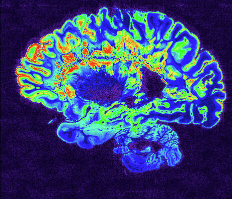
What do hiccups have to do with brain development?
Sambhavi Sneha Kumar explores the origins of hiccups in cerebral development

Perhaps one of the most intricate processes science can explore is that of the development of an embryo in utero to a functional adult. The Roman philosopher Seneca once proposed that a human embryo is simply an ‘adult in miniature’, faced only with the task of increasing in size. Of course, it is now apparent that this is not the case - the complexity and structure of a newborn baby’s organ systems is entirely different to that of the primitive embryo, and continues to change as life progresses. One organ in which this is particularly notable is the brain.
Neuroplasticity is defined as the ability of the brain to form and reorganise connections between its synapses (minute gaps connecting neurons), typically either in response to learning or traumatic injury. The principle usually invoked to explain this is Hebb’s rule, which states that repeated stimulation of a postsynaptic cell by a particular presynaptic cell, increases the firing efficiency of this synapse. While the brain develops its structure before birth, it continues to remodel itself throughout life, notably during the maturation phase of an organism. Neural connections for different brain functions develop sequentially, with the development of sensory pathways in humans peaking at around 4 months of age, language pathways at around 9 months and higher cognitive functions at around 7 years.
Experiments have been carried out to demonstrate that raising animals in deprived environments (for example, darkness, silence or social isolation) can cause defects in brain development and inhibit sensory function. Already in 1978, a study showed that when one eyelid for a kitten was sutured closed, the kitten experienced subsequent vision loss. This prompts the question as to whether it is possible to permanently enhance motor and sensory functioning by changing the stimulating environment of a newborn.
Babies born prematurely tend to be prone to more frequent hiccuping
The plasticity of the brain is at its maximum during what are known as ‘critical periods’, which are maturation times where some crucial experience will have its maximum effect on brain development. If an organism does not undergo the particular experience at the correct time or undergoes it at some later time, the developmental effect may be suboptimal. For example, in babies with congenital deafness, a cochlear implant should be placed within the first 7 years of life to ensure maximum efficacy. While the end of the critical period does not mean a complete loss of synaptic plasticity, the plasticity becomes more limited to synapses confined within a small area and therefore the effect may be less significant and more difficult to achieve.
There are several examples where neuroplasticity can give rise to different outcomes, ranging from recovery from brain injuries during the time periods surrounding birth to the establishment of essential life functions such as breathing.
While strokes in babies are extremely rare, they can occur, for example due to the formation of abnormal clots. A stroke to one side of the brain has different implications for newborns than for adults due to the increased plasticity of the developing brain. A stroke affecting the left hemisphere of the brain can lead to long-lasting language defects in adults. In infants however, while there may be transient delays in the production and understanding of language, normal linguistic development is typically achieved by school age. It has been hypothesised that neuroplasticity can allow for the role of speech and language processing to be shifted to the right side of the brain that was unaffected by the stroke.
Hiccups are one of the
earliest established patterns of activity
Recent research into brain activity pattern using electroencephalograms has implicated that the action of a hiccup may have some evolutionary significance, rather than simply being an involuntary contraction of the diaphragm caused by irritation of the phrenic nerve that supplies it. Hiccups are one of the earliest established patterns of activity, occurring in the fetus in the uterus as early as nine weeks. The study found that the diaphragm muscles causing the hiccup resulted in two large brainwaves on the electroencephalogram followed by a smaller third brainwave that appeared similar to those caused by an auditory stimulus. Researchers believe that this could be an example of how processing multi-sensory inputs allows for the development of brain connections - a newborn baby may be able to connect the ‘hic’ sound of hiccup with the sensation of diaphragm contraction and be able to subsequently utilise voluntary diaphragm contraction in breathing. This may be a remnant from amphibian respiration, in which gills are used to gulp air via a similar motor pathway that is used in hiccuping.
Interestingly, babies born prematurely tend to be prone to more frequent hiccupping, supporting this theory as phenomenon that can have its effect throughout life, its role in brain development in the infant stages of life is particularly significant.
Previous research had hypothesised that hiccups evolved as a reflex to aid the clearance of air from the stomach, with air trapped in the stomach of suckling newborns stimulating the sensory nerves in the stomach. This suggestion arises from the fact that apparently only milk-drinking animals actually have hiccups. While the exact mechanisms are not yet fully understood, imaging techniques such as functional MRI and electroencephalograms may shed light on variations in regional function of the brain that arise as a consequence of neuroplasticity.
 News / Local business in trademark battle with Uni over use of ‘Cambridge’17 January 2026
 News / Cambridge bus strikes continue into new year16 January 2026
 News / News in Brief: cosmic connections, celebrity chefs, and ice-cold competition18 January 2026
 Comment / Fine, you’re more stressed than I am – you win?18 January 2026
 Film & TV / Anticipating Christopher Nolan’s The Odyssey17 January 2026


