The microscope shining a light on the building blocks of life
Laura Shen examines the role of the cryo-electron microsope in today’s scientific world and how its need was realised
When Antonie van Leeuwenhoek first looked though a microscope, he could barely believe the existence of trillions of microorganisms which populate our world. Curiosity has driven us further – to try to explain everything, and that which could be seen by naked eyes can no longer offer sufficient explanations. The invention of the optical microscope had marked a new era in biology and revolutionized the way people viewed the world. Today, we are on the frontier of a similar revolution, spearheaded by the cryo-electron microscope.
As we saw with the first microscopes, life is made from miniature building blocks, or cells – now understood as an essential division of life. Since we are built from cells, perhaps we should ask: what are cells made of? Cryo-electron microscopy (cryo-EM) answers questions on this subcellular scale. Lipids (fats), proteins, nucleic acids and sugars comprise the organic products that populate all living cells, along with various salts and small molecules.
These products combine and recombine in billions of permutations as they work to sustain life. Some molecules are drivers, ferrying proteins and ions across cell boundaries. Some are chefs, feeding the hungry with electrons full of energy. Others are managers, making sure every molecule is on task, and recruiting help when necessary. The cell is far more than a well-oiled machine; it is a bustling metropolis. With the invention of cryo-EM, we finally shed light on the microscopic alien life inside.
As cellular components are incredibly small, studying them is no doubt a daunting task. Previous techniques relied on large quantities of high purity molecules packed in an organized lattice for structural determination, but such techniques limit the molecules’ mobility. As molecular functions are synchronous with structures and movements, accurate recordings of both are necessary for complete understanding.
Cryo-EM, by rapidly freezing molecules in the midst of their activity and imaging them in their various frozen states, can more completely capture the molecules’ range of motion. The information they reveal points to a directed and purposeful activity within the cell. Only by understanding the cell’s individual components can we fully elucidate how cells function.
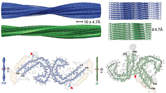
Macromolecular malfunction underlies the majority of non-infectious disease. In July 2017, cryo-EM revealed the first images of tau polymers. Tau proteins have long been hypothesized to form polymers in the brain, triggering neuron death, manifesting as Alzheimer’s disease.
Previously, imaging these molecules would have been impossible. Now, armed with a new method of inquiry, they may be directly investigated and targeted for drug design. In the same manner, cryo-EM may shed light on the multitides of unknown processes our bodies still hide. What makes the immune system attack harmless allergens? How do cancers evade drugs? How are genes turned on and off? With the advent of this new age, whys and wherefores are not from from the horizon.
 News / Simon Goldhill resigns following sexual misconduct investigation16 June 2026
 Comment / Exercise, academia, and the Cambridge addiction to both17 June 2026
 Theatre / A celebration of Queer Love in sacred spaces: Romeo and Juliet in Peterhouse Chapel16 June 2026
 Music / ‘Returning again and again’ to the Bateman room16 June 2026
 Lifestyle / Breaking no contact…hours15 June 2026
 Lifestyle / You can’t always get what you want 18 June 2026
 Theatre / ‘Mental illness doesn’t exist in a vacuum’ says Obsessive, Compulsive, Divine 18 June 2026
 News / Caius to begin antisemitism inquiry after Nazi symbol carved into bench18 June 2026
 Film & TV / Gen Z, FilmTok and the A24 problem17 June 2026
 Features / Are the Footlights getting dimmer?17 June 2026