


How a Cambridge lab created an artificial embryo
The ground-breaking stem cell research could transform our understanding of early embryonic development
Science is one crucial step closer to creating mouse embryos in a Petri dish. Researchers at the Department of Physiology, Development and Neuroscience have successfully developed an artificial environment for mouse stem cells to self-organise into embryo-like structures.
Their findings are likely to offer unprecedented insight into early mammalian development, shedding light on why so many human pregnancies fail during that time.
“Getting this to work required lots of patience, many trials and inspired guesswork,” said Professor Magdalena Zernicka-Goetz, principal researcher on the project.
The breakthrough, published in Nature Cell Biology last week, is that the embryo-like structures grown in the lab can now develop in a condition mirroring that of the earliest stages of life. In the absence of any jelly scaffolding, this embryonic growth comes purely from stem cells.
Up till now, researchers needed to add 3D jelly-scaffolding to artificially grow embryo-like structures in a Petri dish, around which the cells could develop.
Professor Zernicka-Goetz’s team discovered that it is possible to replace the jelly with the extracellular matrix produced by the primitive endoderm stem cells themselves.
This procedure could be crucial in accelerating the pursuit of vital answers to some of the most important questions of early life.
In mammals, once an egg is fertilised by a sperm cell, it divides multiple times to generate a small, free-floating ball - called the blastocyst - comprising three types of stem cells.
“Getting this to work required lots of patience, many trials and inspired guesswork.”
The first type is called embryonic stem cells. They have been called "Building blocks of life", because they will later demonstrate the remarkable ability to develop into many different cell types, from lung tissue to neurons.
The other two stem cell types create the right environment for the embryo to develop into a foetus: trophoblast stem cells, which will go on to form the placenta, and primitive endoderm stem cells which will form the yolk sac, to ensure that the foetus’ organs develop properly and to provide essential nutrients.
Only in the presence of these three stem cells will a process occur that biologist Lewis Wolpert has called “the truly most important time in your life”: gastrulation. Here, the cells undergo large-scale reorganisation and the embryo transforms from being a single layer to three layers: an inner layer (endoderm), middle layer (mesoderm) and outer layer (ectoderm), determining the tissues or organs into which the cells will then develop.

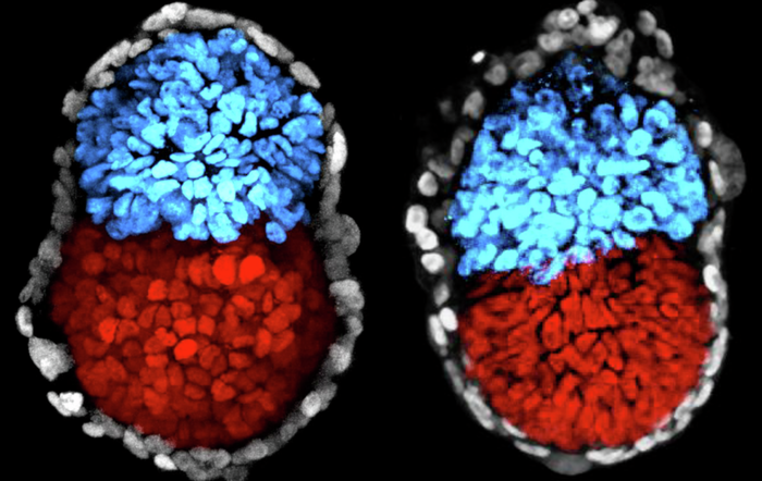
With all three stem cell types – and nothing else – present in the culture dish, the researchers were, for the first time, able to watch an artificially created embryo gastrulate. “Seeing these multiple embryo-like structures growing in a culture dish was incredible; we simply marvelled at life and the ability of these cells organise themselves into proper structures”, says Professor Zernicka-Goetz.
Under the microscope, the researchers watched their mouse-models grow into the equivalent of a 16-day-old human embryo. Looking ahead, the lab hopes to plant their embryos into foster mouse mothers.
The findings will allow the team to explore how embryos develop following implantation – a period of life which is otherwise impossible, and illegal, to explore.
In order for researchers to study human embryos, people who undergo in-vitro fertilisation sometimes donate embryos which were not implanted back into the mother’s womb to science. According to internationally agreed ethics guidelines (and UK law), however, these embryos have to be destroyed after 14 days, well before the embryo develops a nervous system which would allow it to feel things. Because of this, very little is known about embryonic development after two weeks.
It is likely that many diseases have their origins in these early phases of development, but how this happens is poorly understood. It also remains unclear why so many pregnancies fail during the first trimester, with approximately ten to twenty percent ending in a miscarriage.
Prof Zernicka-Goetz’s new embryos will allow scientists to tackle these questions and explore what has, until now, been a black box: the first few weeks and months of mammalian life.
Reference
Sozen, B et al. Self-assembly of embryonic and two extra-embryonic stem cell types into gastrulating embryo structures. Nature Cell Biology; 23 Jul 2018; DOI: 10.1038/s41556-018-0147-7
 News / Night Climbers call for Cambridge to cut ties with Israel in new stunt15 April 2024
 News / Police to stop searching for stolen Fitzwilliam jade17 April 2024
 News / Cambridge University cancer hospital opposed by environmental agency12 April 2024
 Interviews / In conversation with Dorothy Byrne1 March 2024
 Features / Cambridge’s first Foundation Year students: where are they now?7 April 2024
 Interviews / ‘It fills you with a sense of awe’: the year abroad experience17 April 2024
 Sport / Kabaddi: the ancient sport which has finally arrived in Cambridge17 April 2024
- News / Police to stop searching for stolen Fitzwilliam jade17 April 2024
 Fashion / The most viral makeup look of 2024 so far16 April 2024
- News / Night Climbers call for Cambridge to cut ties with Israel in new stunt15 April 2024